


Integrated Management of Endo and Exo-pelvic Sacrococcygeal Teratomas (Altman type III): Report of Two Cases


Abstract
Sacrococcygeal teratoma (SCT) is the most common congenital germ cell tumor in neonates and infants, originating from pluripotent cells of the primitive streak. It may present with external, internal, or mixed pelvic components according to the Altman classification. Although advances in prenatal ultrasonography and fetal magnetic resonance imaging have improved early detection and perinatal management, delayed presentation remains frequent in low-resource settings, often resulting in large and anatomically complex tumors.
We report two cases of sacrococcygeal teratoma with combined endopelvic and exopelvic extension managed in tertiary pediatric surgical units in Benin.
The first case involved a 15-month-old girl presenting with a neglected genitorectal mass evolving since infancy. Imaging revealed a type III SCT with a predominant intrapelvic component. Histopathological examination confirmed an immature teratoma.
The second patient presented with a similar tumor exhibiting both external and intrapelvic extension, posing significant surgical challenges.
In both cases, treatment consisted of complete tumor excision associated with coccygectomy, which remains essential to reduce the risk of recurrence. Pre and postoperative management required a multidisciplinary approach involving pediatric surgeons, anesthesiologists, radiologists, and oncologists to ensure optimal follow-up and oncological surveillance.
These cases highlight the diagnostic and therapeutic challenges of SCT in resource-limited settings. Early diagnosis, precise radiological assessment, and radical surgical resection are critical to improving outcomes and minimizing recurrence and malignant transformation.
Keywords: Sacrococcygeal teratoma; Pediatric surgery; Coccygectomy
Introduction
Sacrococcygeal teratoma (SCT) is the most common congenital germ cell tumor in the fetus and newborn, with an estimated incidence ranging from 1 in 35,000 to 1 in 40,000 live births 1,2. These tumors originate from pluripotent remnants of the primitive streak and Hensen node, which explains their ability to differentiate into tissues derived from the three germ layers, and its development may be entirely external, internal, or mixed, as defined by the Altman classification 1,2,3,4.
This classification remains clinically relevant because it correlates with operative difficulty and risk of pelvic organ compression. Type III lesions are characterized by a predominantly pelvic mass with a smaller external component and may present with constipation, urinary retention, infection, or progressive perineal swelling when diagnosis is delayed 4, 5.
Advances in imaging techniques, particularly prenatal ultrasonography and magnetic resonance imaging (MRI), have significantly improved antenatal diagnosis, enabling early identification of potential complications such as fetal hydrops and facilitating therapeutic decision-making6. Postnatal CT or MRI remains essential to evaluate intrapelvic extension, tumor composition, relation to pelvic organs, sacral anatomy, and surgical resectability6,7.
Surgical management remains the gold standard and consists of complete tumor excision combined with coccygectomy, which is essential to significantly reduce the risk of recurrence reported in the literature 8,9. Long-term surveillance is recommended because recurrence may occur even after apparently complete resection 9.
For tumors containing immature or malignant germ cell elements, multidisciplinary management involving pediatric oncology is required. International cooperative groups such as the Malignant Germ Cell International Consortium (MaGIC) and the Children’s Oncology Group (COG) have refined risk-adapted approaches integrating surgery, tumor markers, and platinum-based chemotherapy 10.
We report two cases of sacrococcygeal teratoma with combined endopelvic and exo-pelvic components, highlighting diagnostic and therapeutic challenges. These cases are discussed in light of recent advances and the specific constraints encountered in low-resource settings.
Cases
Case (1)
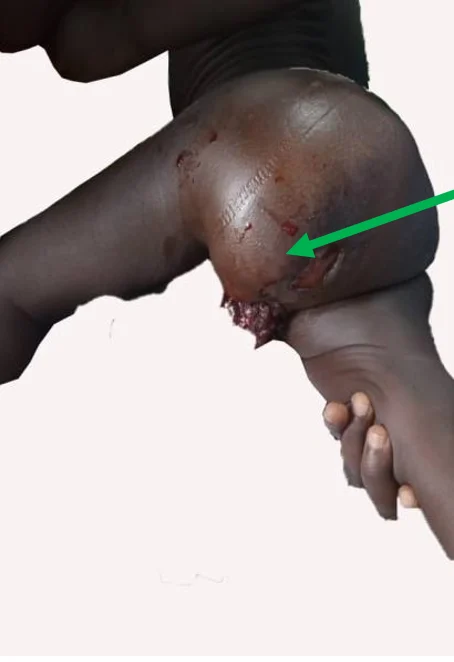
A 15-month-old girl was referred for a large genitorectal mass suggestive of a delayed presentation of sacrococcygeal teratoma (Figure 1). We find no prenatal diagnosis and no maternal complication. Born in a peripheral hospital at term after an uncomplicated pregnancy and vaginal delivery. She was admitted to our hospital due to the progression of the swelling, which included a weeping wound at the tip. There was no sacrococcygeal swelling at birth.
Figure 1. Large exo-pelvic sacrococcygeal mass
The onset appears to have occurred three months prior to admission, with the development of a swelling in the sacrococcygeal region. This required several consultations at peripheral clinics and unsuccessful traditional treatments. Occasional constipation and urinary retention two days before the consultation, which prompted the insertion of a urethral catheter upon admission. No other associated pathology.
A biopsy performed on the perineal mass revealed fibro-adipose and striated muscle tissue infiltrated by a tumor proliferation composed of monotonous round cells, occasionally nucleolated, arranged in rosettes and micropapillary structures. An associated inflammatory infiltrate rich in neutrophils was also observed.
Immunohistochemical analysis demonstrated expression of alpha-fetoprotein (AFP) by tumor cells, with no expression of desmin or myogenin. These findings raised suspicion for a malignant yolk sac component within a mixed germ cell tumor rather than pure immature teratoma alone.
Computed tomography (CT) imaging showed a type III sacrococcygeal teratoma according to the Altman classification. The lesion demonstrated a large presacral component extending anteriorly toward the bladder neck, posterior vaginal plane, and distal rectum, making primary resection technically hazardous because of the risk of incomplete margins, hemorrhage, and pelvic nerve injury (Figure 2A). Serum AFP level was 10 ng/mL, within the normal range for age.
Given the infiltrative involvement of the genitourinary region and histological findings, a multidisciplinary team decision was made to initiate neoadjuvant chemotherapy consisting of four cycles of the VIP regimen (etoposide, Ifosfamide, and cisplatin), prior to surgical management.
Although immature teratoma is not highly chemosensitive, chemotherapy was justified because of the suspected AFP-positive malignant germ cell component and the locally advanced unresectable pelvic extension at presentation.
After two months of chemotherapy, a significant reduction in the endo and exo-pelvic component was observed (Figure 2B). Imaging (CT scan) showed better separation between the bladder neck and rectal wall, decreased pelvic occupancy, and clearer presacral planes, improving resectability.
Figure 2. Complete excision of both components via a posterior approach including coccygectomy after neoadjuvant chemotherapy
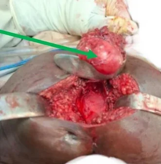
Figure 2A. Green arrow: Exo-pelvic mass
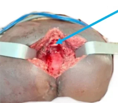
Figure 2B. Blue arrow: sacrum
Complete surgical excision of the residual tumor was then performed by a posterior approach, including a coccygectomy. The endopelvic component was removed after a deep and anatomical presacral dissection, pelvic autonomic nerves were visually preserved, and no macroscopic invasion of the pelvic splanchnic plexus was identified after tumor regression (Figure 2).
Figure 3. 3A = Initial CT scan, endopelvic and exopelvic mass 3B = CT scan after 2 months of chemotherapy: reduction in the endo and exopelvic component
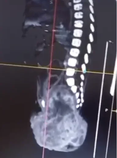
Figure 3A. Initial CT scan, endopelvic and exopelvic mass
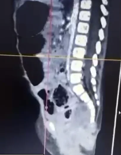
Figure 3B. CT scan after 2 months of chemotherapy
Postoperative course was complicated by a local infection with partial wound dehiscence, with subsequent favorable evolution under appropriate management.
Follow-up included quarterly AFP monitoring, which remained within normal limits. At 20 months follow-up, the patient showed a favorable outcome, with no sphincter or urinary dysfunction and satisfactory wound healing.
Case (2)
An 18-month-old girl was referred for a suppurative extra-pelvic mass with delayed presentation, previously operated on in a peripheral health center without coccygectomy or histopathological analysis. No prenatal diagnosis was made and no maternal complication. Born at term in a peripheral hospital after an uncomplicated pregnancy and vaginal delivery, a small swelling, approximately 2 cm in its longest dimension, has been noted since birth, progressively increasing in size over time.
Clinical examination revealed a distended bladder (urinary retention) associated with suppuration of the posterior surgical wound.
Computed tomography (CT) demonstrated a mass with both intrapelvic and extra-pelvic components, classified as type III according to the Altman classification, with persistence of the coccyx. The lesion contained fatty and calcified elements and caused rectal compression (Figure 4). No metastases were identified.
Figure 4. CT scan = endopelvic and exopelvic mass (Altman type III)
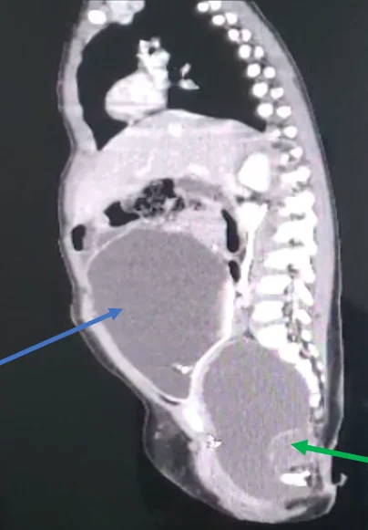
Endopelvic and exopelvic mass (Altman type III) containing fatty and calcified elements (green arrow) compressing the rectum, with persistence of the coccyx. Bladder distension due to compression of the bladder neck (blue arrow)
Serum alpha-fetoprotein (AFP) level was 5 ng/mL, within the normal range for age.
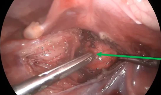
Following multidisciplinary team discussion, complete surgical excision was indicated. Management included a laparoscopic approach for mobilization of the endopelvic component (Figure 5), followed by a posterior approach allowing complete excision of both components along with coccygectomy.
Figure 5. Laparoscopic dissection and mobilization of the endopelvic component of the sacrococcygeal teratoma
Histopathological examination revealed a fibrocongestive cystic wall lined by keratinizing squamous epithelium, containing multiple differentiated tissue elements (adipose, respiratory, cartilaginous, and osseous), with no immature component. Complete resection was confirmed.
The postoperative course was uneventful. Follow-up included quarterly AFP monitoring, which remained within normal limits.
At 9 months follow-up, the short outcome was favorable, with no clinical or biological evidence of recurrence. there was no short-term clinical or biological evidence of recurrence; however, longer surveillance remains necessary before concluding a durable favorable outcome.
Table 1. Comparison between Case (1) & (2)
| Variables | Case (1) | Case (2) |
|---|---|---|
| Sex | Female | Female |
| Age at presentation | 15 months | 18 months |
| Birth History | Term birth; no prenatal diagnosis | Term birth; no prenatal diagnosis |
| Pregnancy history | No maternal complication | No maternal complication |
| Time symptoms first noted | Since infancy, progressive mass enlargement | Neonatal mass, incompletely treated |
| Associated disease | Occasional constipation, urinary retention + wound suppuration | Urinary retention + wound suppuration |
| Altman type | III | III |
| Main pelvic effects | Compression of genitourinary region | Rectal compression + bladder neck compression |
| Histology | Immature teratoma with suspected yolk sac component (AFP) | Mature teratoma |
| AFP preoperative | 10ng/mL | 5ng/mL |
| AFP postoperative | Normal during follow-up | Normal during follow-up |
| Initial treatment | Neoadjuvant VIP chemotherapy | Surgery |
| Surgical approach | Posterior + presacral dissection + coccygectomy | Laparoscopy + posterior approach + coccygectomy |
| Follow-up | 20 months | 9 months |
| Current status | No recurrence, no dysfunction | No early recurrence; longer follow-up needed |
Discussion
Sacrococcygeal teratomas (SCTs) originate from the presacral region and are consistently attached to the coccyx, which justifies systematic coccygectomy during surgical management 1,2. Their marked morphological heterogeneity and variable malignant potential necessitate a rigorous multidisciplinary approach 3.
Although many lesions are detected before birth, delayed presentation persists in low- and middle-income countries due to limited antenatal screening, delayed referral routes, and limited access to pediatric surgical care 1,11. Our two observations, which did not even have an antenatal diagnosis, illustrate this persistent disparity in access to specialized treatments.
The Altman classification remains a key tool for guiding therapeutic strategy, particularly in types II and III characterized by significant intrapelvic extension 4. As in Case 1, such anatomy can lead to compression of the rectum, bladder outlet, ureters, or pelvic neural structures, thus explaining constipation, urinary retention, or progressive perineal swelling. It provides essential anatomical guidance for surgical planning and prognosis 5,8. In Case 2, bladder distension and rectal compression were directly related to the mass effect of the pelvic component.
Advances in imaging have significantly improved the diagnostic approach. Prenatal ultrasonography enables early detection, while fetal magnetic resonance imaging (MRI) provides detailed information on anatomical relationships and tumor composition 6. In selected severe or highly vascularized cases, prenatal interventions may be considered in specialized centers 6, 12.
In resource-limited settings where MRI is not consistently available, CT-Scan often remains the practical modality despite radiation exposure. This was the case in our institution, where CT- Scan was essential for preoperative mapping.
However, complete surgical excision combined with coccygectomy remains the cornerstone of treatment. Recurrence rates, reported to reach 30–40% in cases of incomplete resection or coccyx preservation, strongly support this approach 13,14,15. Long-term follow-up is essential due to the risk of recurrence and late malignant transformation 14. The second case in our series clearly illustrates the consequences of inadequate initial management: the child had undergone prior incomplete surgery in a peripheral center without coccygectomy and later presented with persistent disease requiring redo surgery. This emphasizes the importance of referral of suspected SCT to experienced pediatric surgical teams.
The role of chemotherapy depends on tumor histology and biology. Mature teratomas are generally managed surgically 16,17. In contrast, tumors containing yolk sac tumor elements or other malignant germ cell components require platinum-based chemotherapy according to pediatric germ cell tumor protocols. Its indications are supported by large pediatric oncology series and international treatment protocols 14,17.
The first case in this series demonstrates the value of neoadjuvant chemotherapy in reducing tumor volume and facilitating surgical resection. Histology showed rosettes and micropapillary structures with AFP-positive immunostaining, raising suspicion of an associated yolk sac component despite normal serum AFP. Such discordance has been described and may occur in focal malignant transformation, low tumor burden, partial necrosis, or sampling 18, 19. VIP was selected over standard PEB (Cisplatin, Etoposide + Bleomycin) because of institutional preference to avoid bleomycin-related pulmonary toxicity in an infant requiring future major surgery and repeated anesthesia exposure, while maintaining platinum-based efficacy 20,21,22.
In low-resource settings, SCT management remains challenging due to limited access to advanced imaging, lack of systematic prenatal diagnosis, shortage of specialized pediatric surgeons, limited pathology services, and difficulties in long-term follow-up, particularly for AFP monitoring. These challenges have been reported in African series, including those by Ndour et al. and Moifo et al. 11,23.
Nevertheless, progressive improvements are being observed through case centralization, implementation of multidisciplinary team meetings, enhancement of technical facilities, and alignment with international treatment protocols. Experiences from specialized centers have shown that adherence to standard management principles comprehensive imaging, complete resection with coccygectomy, and prolonged AFP surveillance can yield favorable outcomes comparable to those reported in high-resource settings 24.
The two cases presented illustrate the dual reality of structural constraints and ongoing improvements in management. They also highlight the contribution of laparoscopy in addressing endopelvic components, facilitating complete tumor excision in selected cases.
These findings underscore the need to strengthen specialized training, improve access to advanced imaging modalities, and promote systematic referral to specialized centers in order to optimize long-term outcomes in patients with SCT.
Conclusion
The management of sacrococcygeal teratomas relies on a rigorous multidisciplinary approach, combining early diagnosis, complete surgical excision including systematic coccygectomy, and prolonged alpha-fetoprotein (AFP) monitoring.
Recent advances in imaging, surgical techniques, and oncological protocols demonstrate that, even in low-resource settings, adherence to international standards can achieve satisfactory outcomes. Strengthening diagnostic and surgical capacities, along with systematic referral to specialized centers, remains essential to improve long-term prognosis.
License
© Author(s) 2026.
This is an open-access article distributed under the terms of the Creative Commons Attribution 4.0 International License (CC BY 4.0), which permits unrestricted use, distribution, and reproduction in any medium, and unrestricted adaptation and reuse, including for commercial purposes, provided you give appropriate credit to the original author(s) and the source, provide a link to the Creative Commons license, and indicate if changes were made.
To view a copy of this license, visit https://creativecommons.org/licenses/by/4.0/.
References
-
Tosun M, Cam İ, Uslu H, Doğan Y, Anık Y. A single-center experience of magnetic resonance imaging findings of fetal sacrococcygeal teratomas. Turk J Med Sci. 2022;52(4):1190-6.
-
Carr S, Safavi A, Skarsgard ED. Case reports: management challenges in pediatric germ cell tumors. Front Pediatr. 2021;9:659083.
-
Lew CZ, Liu HC, Hou JY, Huang TH, Yeh TC. Pediatric extracranial germ cell tumors: review of clinics and perspectives in application of autologous stem cell transplantation. Cancers (Basel). 2023;15(7):1998.
-
Altman RP, Randolph JG, Lilly JR. Sacrococcygeal teratoma: a survey. J Pediatr Surg. 1974;9(3):389-98.
-
Salim A, Raitio A, Losty PD. Long-term functional outcomes of sacrococcygeal teratoma – a systematic review of published studies exploring ‘real world’ outcomes. Eur J Surg Oncol. 2023;49(1):16-20.
-
Li X, Lin H, Hu K, Peng Y. Prenatal MRI diagnosis and outcomes of abdominal or sacrococcygeal teratomas and parasitic fetuses. Front Pediatr. 2023;11:1181110.
-
Mihai BM, Bohiltea RE, Dima V, Veduta A, Salmen T, Georgescu TA, et al. Update in the prenatal management of sacrococcygeal teratomas and the outcome of the newborn – case report. Ro J Pediatr. 2021;70(3):273-7.
-
Gabra HO, Jesudason EC, McDowell HP, Pizer BL, Losty PD. Sacrococcygeal teratoma–a 25-year experience in a UK regional center. J Pediatr Surg. 2006;41(9):1513-6.
-
Elgendy A, AbouZeid AA, El-Debeiky M, Mostafa M, Takrouney MH, Abouheba M, et al. Management strategy and outcomes of sacrococcygeal teratoma – an Egyptian multicenter experience. World J Surg Oncol. 2023;21(1):294.
-
Pashankar F, Murray MJ, Gell J, MacDonald N, Shamash J, Billmire DF, et al. Consensus and controversy in the management of paediatric and adult patients with ovarian immature teratoma: the Malignant Germ Cell International Consortium perspective. eClinicalMedicine. 2024;69:102453.
-
Ndour O, Faye PA, Alumeti D, Ndoye MD, Ngom G. Sacrococcygeal teratoma in sub-Saharan Africa: management challenges. Afr J Paediatr Surg. 2021;18(2):101-6.
-
Abiad M, Zargarzadeh N, Javinani A, Krispin E, Shamshirsaz AA. Fetal teratomas: advances in diagnosis and management. J Clin Med. 2024;13(20):6245.
-
De Backer A, Madern GC, Hakvoort-Cammel FG, et al. [Study of factors associated with recurrence in children with sacrococcygeal teratoma]. J Pediatr Surg. 2006;41(1):173-81
-
Padilla BE, Vu L, Lee H, MacKenzie T, Bratton B, O’Day M, et al. Sacrococcygeal teratoma: late recurrence warrants long-term surveillance. Pediatr Surg Int. 2017;33(11):1189-94.
-
Rescorla FJ. Pediatric germ cell tumors. Semin Surg Oncol. 1999;16(2):144-58.
-
Bode PK, Blasco-Santana L, Colmenero I, Reyes-Múgica M. Germ cell tumors in children. Virchows Arch. 2025;486(1):65-79.
-
Bhuta R, Shah R, Gell JJ, Poynter JN, Bagrodia A, Dicken BJ, et al. Children’s Oncology Group’s 2023 blueprint for research: Germ cell tumors. Pediatr Blood Cancer. 2023;70 Suppl 6:e30562.
-
Jezierska M, Gawrychowska A, Stefanowicz J. Diagnostic, prognostic and predictive markers in pediatric germ cell tumors–past, present and future. Diagnostics (Basel). 2022;12(2):278.
-
Lobo J, Leão R, Jerónimo C, Henrique R. Liquid biopsies in the clinical management of germ cell tumor patients: state-of-the-art and future directions. Int J Mol Sci. 2021;22(5):2654.
-
Jayakrishnan B, Kausalya R, Al-Rashdi HA, Davis K, Ali J, Al-Harthy M, et al. Bleomycin and perioperative care: a case report. Sarcoidosis Vasc Diffuse Lung Dis. 2023;40(3):e2023030.
-
Farazdaghi A, Vaughn DJ, Singhal S. Pulmonary metastasectomy for germ cell tumors. Ann Thorac Cardiovasc Surg. 2019;25(6):289-95.
-
Rescorla F, Billmire D, Stolar C, et al. The effect of cisplatin dose and surgical resection in children with malignant germ cell tumors at the sacrococcygeal region: a pediatric intergroup trial (POG 9049/CCG 8882). J Pediatr Surg. 2001;36(1):12-7.
-
Moifo B, Mouafo Tambo FF, Nguefack S, Sando Z, Abogo S, Gonsu Fostin J. [Teratome sacro-coccygien vu tard: aspects cliniques, radiologiques et thérapeutiques à propos de deux cas]. Mali Med. 2014;29(1):53-7.
-
Srivatsa S, Gil L, Zhang Y, et al. Optimizing surveillance strategies for sacrococcygeal teratoma: A Midwest pediatric surgery consortium multi-institutional study. Int J Cancer. 2025;157(10):2103-13.
How to Cite
Gildas YM, Alain BGM, Max MN, Géraud SS, Romeo MSE, Isaac B, et al. Integrated Management of Endo and Exo-pelvic Sacrococcygeal Teratomas (Altman type III): Report of Two Cases. OncoDaily Med J. 2026;3(2). doi:10.69690/ODMJ-011-0826-7849