


Ecthyma Gangrenosum in a Child with Acute Lymphoblastic Leukaemia
Abstract
Introduction: A 12-year-old female child presented with a progressive skin lesion diagnosed as Ecthyma gangrenosum, the growth of Pseudomonas aeruginosa was confirmed based on lesion progression, positive blood, patient’s immunocompromised status, and tissue biopsy. The lesion was managed and treated with intravenous antibiotics and antifungals, conservatively with daily
dressing.
Case presentation: A 12-year-old female, on relapsed leukemia protocol. She was admitted to the ICU with a disturbed consciousness level, hypotension, and hemorrhagic skin lesions in both the upper and lower limbs. Her medical history included a one-week duration of severe bone pain, fever, rhinorrhea, cough, epistaxis, and gastrointestinal symptoms. Initial investigations revealed elevated inflammatory markers, abnormal liver and kidney function, and a positive blood culture for Pseudomonas aeruginosa. Started supportive treatment and an empirical; HD Meronum, Amikin, Vancomycin, and Ambizome.
After 48 hours she was afebrile and vitally stable and she was transferred to a pediatric ward. Despite this improvement, new progressive skin lesions with hemorrhagic necrosis developed, and the voriconazole level was within normal range. Blood culture sensitivity results led to the addition of intravenous ciprofloxacin, and a tissue biopsy confirmed the presence of Pseudomonas aeruginosa, a high-level AmpC producer. Gentamicin was added to her regimen. After two weeks, her skin lesions improved significantly, and her blood counts and CRP returned to normal. She was discharged after 25 days with oral antibiotics (Co-trimoxazole and Ciprofloxacin) and follow-up recommendations.
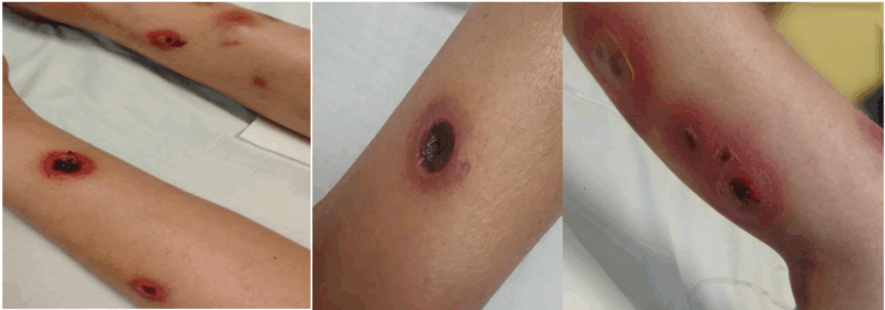
Figure: Ecthyma gangrenosum caused by P. aeruginosa
Ecthyma gangrenosum (EG) is a rare cutaneous infection that mostly occurs in immunosuppression patients with considerable morbidity and mortality, caused by bacteria, fungi, and virus infection. P. aeruginosa is a rapidly spreading single or multiple necrotic skin lesion that might involve one or more sites in the
body.
The diagnosis is based on skin biopsy and blood cultures. A multidisciplinary team should be considered to improve outcomes. Combined antimicrobial and surgical debridement should be considered as a standard of care, medical therapy alone may be sufficient for some patients. The clinical response depends on factors such as underlying diseases, severity and type of infection, adequate source control, and response to antibiotics. Local antiseptic agents like citric acid are recommended due to their antibacterial properties and role in enhancing wound healing.
Conclusion: EG is an infectious complication described in pediatric patients under treatment for hematological malignancy. Prompt recognition and timely adequate treatment are crucial for the prognosis.