


Posterior Fossa Ependymoma in a Child with Neurofibromatosis Type 1: A Rare Association
Posterior Fossa Ependymoma in a Child with Neurofibromatosis Type 1: A Rare Association
Introduction
Ependymomas accounts for 6-12% of pediatric intracranial tumors, they are glial tumors commonly found in the posterior fossa of children and are typically associated with NF2. Their occurrence in NF1 is exceptionally rare. Neurofibromatosis type 1 (NF1) is a distinct autosomal dominant disorder characterized by café-au-lait macules, Lisch nodules, osseous lesions, and predisposition to optic pathway gliomas and other astrocytomas. We report a unique case of posterior fossa ependymoma in a child with NF1. The objective of the study was to describe the clinical presentation, diagnosis, and management of a pediatric NF1 patient who developed a posterior fossa ependymoma.
Case Presentation
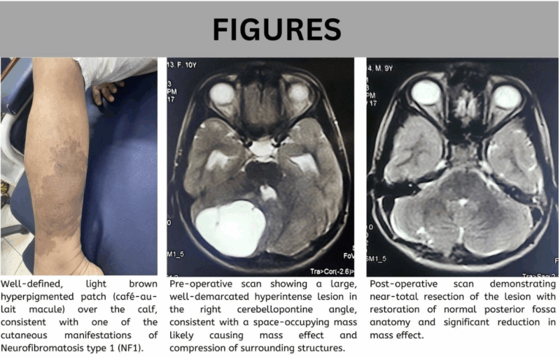
A 10-year-old girl with a known family history of Neurofibromatosis Type 1 (NF1) presented with a 4–5-month history of progressively worsening early-morning holocranial headaches, frequent vomiting, and cerebellar signs including gait imbalance and dysdiadochokinesia. MRI of the brain revealed a well-defined, contrast-enhancing mass in the fourth ventricle, compressing the brainstem and causing obstructive hydrocephalus. She underwent gross-total surgical resection at a tertiary pediatric hospital in Karachi, Pakistan. Histopathological examination confirmed a WHO Grade II ependymoma, showing perivascular pseudorosettes and moderate nuclear pleomorphism. Postoperative cerebrospinal fluid cytology was negative for malignant cells. Following surgery, detailed physical examination revealed multiple NF1 stigmata: over six café-au-lait macules (>5 mm), bilateral Lisch nodules on slit-lamp examination, anterior tibial bowing, and a first-degree relative with similar features. These findings met four NIH diagnostic criteria, confirming a clinical diagnosis of NF1. No molecular testing was performed. The patient recovered without neurological deficits and was referred for oncology follow-up. This is a rare instance of intracranial ependymoma in an NF1 patient, an association typically observed in NF2, and possibly the first reported case involving the posterior fossa in a child with NF1.
Conclusion
This case highlights an exceptionally rare occurrence of a posterior fossa ependymoma in a child with NF1, thus, expanding the known phenotypic spectrum of NF1 and suggesting the long-term neuroimaging surveillance that may be considered in such patients.
Conflict of Interest: None
Funding: None
Disclosure statement: None
License: This article is published under the terms of the Creative Commons Attribution 4.0 International License (CC BY 4.0).
©️ Muhammad Kashan 2025. This license permits unrestricted use, distribution, and reproduction in any medium, provided the original author and source are credited.